Anatomy and physiology is the scientific foundation on which every clinical decision in healthcare ultimately rests. Before a nurse can understand why a patient in heart failure retains fluid, they must understand how the kidneys regulate sodium and water balance. Before a respiratory therapist can interpret an arterial blood gas, they must understand how the lungs exchange gases across the alveolar membrane. Before a physical therapist can design a rehabilitation program following a stroke, they must understand which neural pathways govern voluntary motor function and where they were disrupted. Before a medical student can reason through a diagnosis of diabetic ketoacidosis, they must understand how the endocrine pancreas regulates glucose metabolism and what happens when that regulation fails. Anatomy and physiology is not a prerequisite to be completed and forgotten. It is the explanatory framework that gives clinical knowledge its depth, coherence, and meaning. Every time a healthcare professional understands not just what to do but why, they are drawing on this foundational science. That is why mastering it thoroughly, rather than simply passing through it, is one of the most consequential investments any healthcare student can make.
The challenge is that the content is vast, interconnected, and conceptually demanding in ways that are unlike most other subjects in a healthcare curriculum. Memorizing the names of bones, muscles, and cranial nerves is only the beginning. The deeper task is understanding how structures relate to one another, how organ systems interact and regulate each other through feedback mechanisms, how disruptions in normal physiology produce the clinical signs and symptoms that healthcare providers must recognize and interpret, and how the microscopic world of cells and tissues gives rise to the macroscopic world of organs and systems. That kind of integrative, applied understanding is not built by passive reading. It is built through active engagement with content that requires the learner to think, apply, connect, and analyze. That is exactly what this test bank is designed to facilitate.
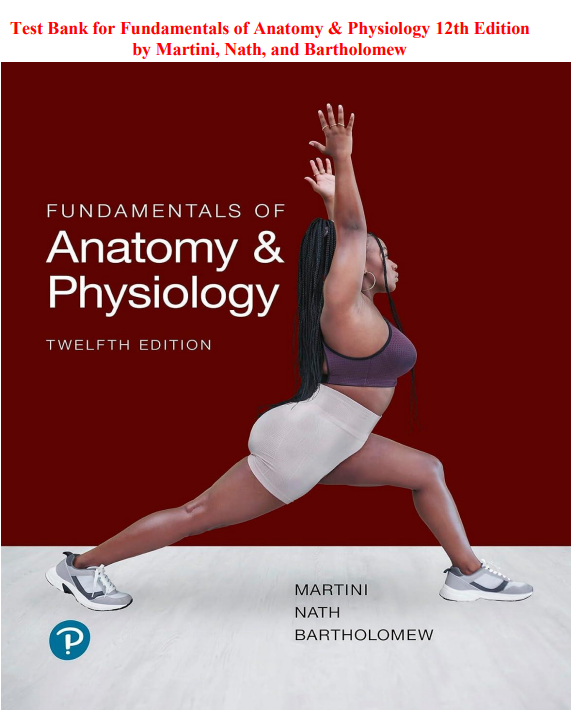
Fundamentals of Anatomy and Physiology by Martini, Nath, and Bartholomew has long been one of the most comprehensive, visually rich, and pedagogically effective anatomy and physiology texts available for students in nursing, allied health, and pre-medical programs. The 12th edition continues that tradition with thoroughly updated content reflecting current physiological research, expanded coverage of clinical applications and homeostatic imbalances, improved integration between anatomical structure and physiological function, and a stronger emphasis on the systems-level thinking that healthcare education demands. This test bank was written to match that standard of depth and clinical relevance at every level.
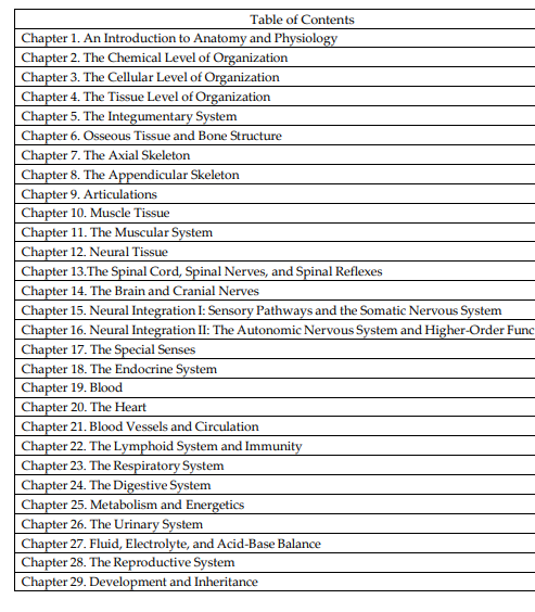
Every question in this resource follows the 12th edition chapter structure. The coverage is exhaustive and carefully organized across the full breadth of the textbook, spanning all eleven organ systems and the foundational sciences that underlie them. Questions address the chemical and cellular foundations of life, including atomic structure, chemical bonds, organic macromolecules, cell structure and organelles, membrane transport mechanisms, cell division, and protein synthesis. Histology content covers the four primary tissue types and their structural and functional characteristics. The integumentary system is covered in depth, including the layers of skin, accessory structures, and thermoregulation. The skeletal system content spans bone tissue structure and remodeling, the axial and appendicular skeleton, articulations and joint types, and the clinical implications of disorders including osteoporosis and fractures. Muscular system content addresses skeletal muscle microstructure, the sliding filament mechanism, neuromuscular junction physiology, muscle fiber types, and smooth and cardiac muscle. Nervous system content covers neuron structure and function, action potential generation and propagation, synaptic transmission, the central and peripheral nervous systems, the autonomic nervous system, and the special senses. The endocrine system is addressed with emphasis on hormone mechanisms, feedback regulation, and the physiological consequences of endocrine disorders. Cardiovascular content covers cardiac anatomy, the cardiac cycle and electrocardiography, hemodynamics, blood pressure regulation, and the properties of blood including hematopoiesis, erythrocyte physiology, hemostasis, and blood typing. Lymphatic and immune system content addresses lymphatic anatomy, innate and adaptive immunity, and the mechanisms of allergic and autoimmune responses. Respiratory system content covers pulmonary anatomy, the mechanics of ventilation, gas exchange and transport, and the regulation of breathing. Digestive system content addresses the anatomy and physiology of each segment of the gastrointestinal tract, the accessory organs of digestion, and the absorption of nutrients. Urinary system content covers nephron structure and function, the processes of filtration, reabsorption, and secretion, urine formation and concentration, and acid-base regulation. Reproductive system content spans male and female reproductive anatomy, gametogenesis, the hormonal control of the reproductive cycle, fertilization, and the physiological changes of pregnancy.
What makes this test bank distinctly valuable is the quality and analytical depth of its answer explanations. Every question presents a concept-based or clinically applied scenario, clearly identifies the correct answer, and follows it with a thorough, well-reasoned rationale. These rationales do not simply confirm what is right. They explain the physiological mechanism or anatomical principle behind the correct choice, address why each of the remaining options is incorrect, and where relevant connect the content to a clinical application or homeostatic imbalance that illustrates why the concept matters in real healthcare practice. That depth of explanation means every question functions as both an assessment and a genuine learning experience. You are not simply practicing answer selection. You are building the integrative physiological understanding that makes the right answer evident under any examination conditions and that will serve you across the entire span of your healthcare education and career.
The file is a fully searchable PDF. When you need to focus your review on renal physiology before a fluid and electrolyte examination, consolidate your understanding of cardiac conduction and electrocardiography before a cardiovascular unit, or drill questions on the mechanisms of hormone action before an endocrine assessment, use Ctrl+F to navigate directly to that content in seconds. No time lost scrolling through chapters you have already mastered. No disruption to your study momentum. Direct, precise access to exactly the content you need at exactly the moment you need it.
Your download link is generated the moment your purchase is confirmed. No waiting period, no account creation, no approval process. Open the file on your laptop, tablet, or phone and begin working through it immediately. The file is yours permanently. No subscription, no renewal fee, and no expiry date.
What You Get
- Full chapter coverage matched to the 12th edition layout across all organ systems and foundational content areas
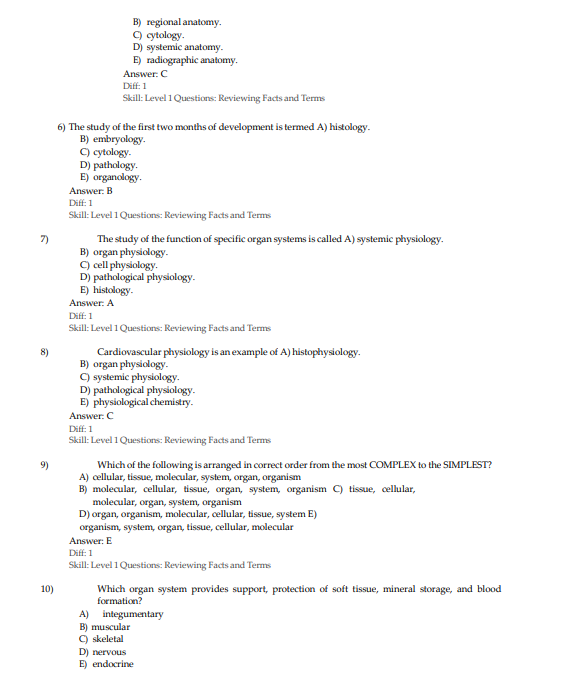
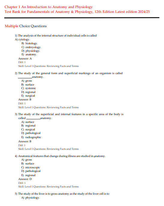
- Concept-based and clinically applied multiple-choice questions reflecting the depth and breadth of the Martini, Nath, and Bartholomew textbook
- Every question answered with the correct response clearly identified
- Detailed rationales explaining the anatomical and physiological reasoning and addressing all answer options
- Searchable PDF format for fast, targeted review by organ system, physiological process, or clinical concept
- One-time purchase with permanent, unlimited file access
Who This Is For
Nursing, allied health, pre-medical, pre-dental, and exercise science students enrolled in introductory or advanced anatomy and physiology courses will benefit most directly from this resource. It mirrors the chapter structure of the Martini, Nath, and Bartholomew textbook closely and is designed to support students as they build the foundational scientific knowledge on which all subsequent healthcare education depends. It is also an excellent preparation tool for students approaching standardized examinations that include anatomy and physiology content, including the HESI A2, TEAS, NCLEX, and various allied health certification examinations. Students who struggled with anatomy and physiology in a previous course and need a structured, explanation-rich resource for review and remediation will find this test bank particularly valuable. Healthcare professionals returning to education after time in practice who need to refresh and consolidate their foundational science knowledge will also benefit significantly from the depth and breadth of this resource.
Sample Questions
Question 1
A physiology student is reviewing the mechanisms by which the kidneys regulate blood pressure. The student learns that when blood pressure drops significantly, specialized cells in the kidney release an enzyme that initiates a hormonal cascade. This cascade ultimately produces a hormone that causes vasoconstriction and stimulates the adrenal cortex to release a mineralocorticoid that promotes sodium and water retention by the kidneys. Which sequence of components correctly describes this pathway in the correct order?
A) Renin released by juxtaglomerular cells → angiotensinogen converted to angiotensin I → angiotensin-converting enzyme converts angiotensin I to angiotensin II → angiotensin II causes vasoconstriction and stimulates aldosterone release from the adrenal cortex → aldosterone promotes sodium and water reabsorption in the distal tubule and collecting duct
B) Aldosterone released by the adrenal medulla → renin converts angiotensinogen to angiotensin II → angiotensin II stimulates antidiuretic hormone release from the posterior pituitary → antidiuretic hormone causes vasoconstriction and promotes water retention in the proximal tubule
C) Erythropoietin released by juxtaglomerular cells → erythropoietin stimulates the liver to produce angiotensinogen → angiotensinogen causes direct vasoconstriction → the adrenal cortex releases cortisol to promote sodium retention in the loop of Henle
D) Renin released by the macula densa → renin directly converts angiotensin I to angiotensin II in the pulmonary circulation → angiotensin II stimulates epinephrine release from the adrenal medulla → epinephrine promotes sodium retention in the proximal convoluted tubule
Correct Answer: A
Detailed Explanation:
The renin-angiotensin-aldosterone system is one of the most physiologically significant and clinically important hormonal regulatory pathways in the human body, and a thorough understanding of its components, sequence, and sites of action is essential for any healthcare student. It is also the basis for an entire class of antihypertensive medications, including ACE inhibitors and angiotensin receptor blockers, which makes understanding this pathway directly clinically relevant.
The sequence begins in the kidney. When arterial blood pressure falls, when blood volume decreases, or when sympathetic nervous system activity increases, specialized smooth muscle cells in the walls of the afferent arterioles of the renal corpuscle, called juxtaglomerular cells or granular cells, respond by releasing the enzyme renin into the bloodstream. Renin acts on a plasma protein called angiotensinogen, which is produced and released by the liver into the general circulation. Renin cleaves angiotensinogen to produce a decapeptide called angiotensin I, which is relatively inactive. As angiotensin I passes through the pulmonary capillaries, it encounters angiotensin-converting enzyme, which is produced in high concentrations by the endothelial cells of the pulmonary vasculature. ACE cleaves two amino acids from angiotensin I to produce the octapeptide angiotensin II, the primary active effector molecule of the system. Angiotensin II exerts two major physiological effects that together restore blood pressure and blood volume: it is a potent vasoconstrictor that increases peripheral vascular resistance directly, and it stimulates the zona glomerulosa of the adrenal cortex to synthesize and release aldosterone. Aldosterone acts on the principal cells of the distal convoluted tubule and collecting duct of the nephron, stimulating the insertion of sodium-potassium ATPase pumps and sodium channels that increase the reabsorption of sodium from the tubular filtrate back into the bloodstream. Water follows osmotically, expanding blood volume and thereby restoring blood pressure. ACE inhibitors work by blocking this conversion step, preventing the formation of angiotensin II and thereby reducing both vasoconstriction and aldosterone-driven fluid retention.
Option B is incorrect because aldosterone is produced by the adrenal cortex, not the adrenal medulla. The adrenal medulla produces catecholamines including epinephrine and norepinephrine. Additionally, renin is an enzyme that acts on angiotensinogen, not on angiotensin II, and it does not perform the conversion described. Option C is incorrect because erythropoietin is a hormone produced by the kidneys that stimulates red blood cell production in the bone marrow. It has no role in the renin-angiotensin-aldosterone system. Option D is incorrect because it contains multiple errors. The macula densa cells are chemoreceptors that monitor the sodium chloride content of the tubular filtrate and signal the juxtaglomerular cells to release renin, but they do not release renin themselves. Additionally, renin does not directly convert angiotensin I to angiotensin II; that conversion requires angiotensin-converting enzyme. Epinephrine is not the mineralocorticoid responsible for sodium retention in this pathway.
Question 2
A healthcare student is studying cardiac physiology and reviewing an electrocardiogram tracing from a patient in a cardiology clinic. The student notes that the PR interval measures 0.28 seconds, which is prolonged beyond the normal range of 0.12 to 0.20 seconds. The QRS complex has a normal duration and morphology. The student is asked to identify which component of the cardiac conduction system is most likely responsible for this delay and to explain the physiological significance of the PR interval. Which explanation is most accurate?
A) The prolonged PR interval reflects delayed ventricular repolarization following contraction, indicating that the bundle of His is conducting too slowly after the ventricles have depolarized
B) The prolonged PR interval reflects slowed conduction through the atrioventricular node, which normally introduces a physiological delay between atrial and ventricular depolarization to allow the ventricles time to fill with blood before contracting
C) The prolonged PR interval reflects delayed sinoatrial node automaticity, indicating that the pacemaker cells are firing less frequently than normal and producing a slower heart rate
D) The prolonged PR interval reflects slowed conduction through the bundle branches, indicating that one or both bundle branches are partially blocked and ventricular depolarization is occurring asynchronously
Correct Answer: B
Detailed Explanation:
The electrocardiogram is a graphical representation of the electrical activity of the heart as detected by surface electrodes, and understanding the physiological correlates of each waveform and interval is a fundamental competency for any healthcare professional working in clinical settings. The PR interval is measured from the beginning of the P wave, which represents atrial depolarization initiated by the sinoatrial node, to the beginning of the QRS complex, which represents the onset of ventricular depolarization. The structures whose conduction times are reflected in the PR interval include the atrioventricular node, the bundle of His, and the proximal bundle branches, but the dominant contributor to the PR interval duration is the atrioventricular node.
The atrioventricular node, located in the inferior interatrial septum near the opening of the coronary sinus, is the only electrical connection between the atria and the ventricles in a normal heart. Its physiological function is to introduce a controlled delay in conduction between atrial depolarization and ventricular depolarization. This delay is essential for coordinated cardiac function because it allows the atria to complete their contraction and deliver their volume of blood into the ventricles through the atrioventricular valves before the ventricles begin to contract. Without this delay, the ventricles might contract before they have received the full atrial contribution to their end-diastolic volume, reducing stroke volume and cardiac efficiency.
The normal PR interval ranges from 0.12 to 0.20 seconds. A PR interval exceeding 0.20 seconds indicates first-degree atrioventricular block, which represents slowed conduction through the atrioventricular node without complete interruption of conduction. Every atrial impulse still reaches the ventricles, but the delay is prolonged beyond the physiological norm. First-degree AV block can be caused by increased vagal tone, certain medications including beta-blockers, calcium channel blockers, and digoxin, electrolyte imbalances, or structural damage to the AV node from ischemia or inflammation.
Option A is incorrect because ventricular repolarization is represented by the T wave on the electrocardiogram, not the PR interval. The PR interval measures the time from the onset of atrial depolarization to the onset of ventricular depolarization, not repolarization events. Option C is incorrect because sinoatrial node automaticity determines the rate of P wave generation and therefore influences the heart rate and the RR interval. Slowed sinoatrial node firing would produce a longer RR interval and a slower heart rate, not a prolonged PR interval. Option D is incorrect because bundle branch conduction is reflected in the duration and morphology of the QRS complex, not the PR interval. A bundle branch block produces a widened QRS complex, typically greater than 0.12 seconds, because ventricular depolarization travels through slower myocardial tissue rather than the specialized conduction system when one bundle branch is blocked.
Question 3
A student in an anatomy and physiology course is studying the process of skeletal muscle contraction at the molecular level. The student understands that muscle contraction requires the interaction of actin and myosin filaments and is regulated by calcium ions and the troponin-tropomyosin complex. The student is asked to arrange the following events in the correct sequence for a single cross-bridge cycle. Which sequence correctly represents the molecular events from the resting state through one complete cross-bridge cycle?
A) Calcium ions bind to troponin C, shifting tropomyosin to expose active sites on actin → myosin head in the cocked position binds to the active site on actin, forming a cross-bridge → power stroke occurs as the myosin head pivots toward the M-line, pulling the actin filament toward the center of the sarcomere → ATP binds to the myosin head, causing it to detach from actin → ATP hydrolysis to ADP and inorganic phosphate re-cocks the myosin head, preparing it for the next cycle
B) ATP hydrolysis occurs first, releasing energy that causes actin to bind to myosin → calcium ions are released from the myosin head to activate troponin → the power stroke occurs before the myosin head detaches from actin → ADP re-cocks the myosin head without requiring calcium → tropomyosin returns to its blocking position before the next cross-bridge cycle
C) Myosin head attaches to actin in the uncocked position → ATP immediately causes the power stroke → calcium is released from the sarcoplasmic reticulum after the power stroke to sustain contraction → ADP and phosphate detach from myosin before the cross-bridge forms → tropomyosin blocks the active sites only after multiple cross-bridge cycles
D) Calcium binds to tropomyosin directly → tropomyosin detaches completely from actin → free actin filaments drift toward the M-line without myosin involvement → myosin binds to actin after the power stroke has occurred → ATP is not required for cross-bridge cycling in resting skeletal muscle
Correct Answer: A
Detailed Explanation:
The sliding filament mechanism of muscle contraction is one of the most conceptually important and frequently examined topics in anatomy and physiology. Understanding the molecular events of cross-bridge cycling requires knowledge of the roles of calcium, troponin, tropomyosin, actin, myosin, and ATP, and the precise sequence in which these molecules interact.
At rest, the myosin-binding sites on the actin thin filaments are physically blocked by the protein tropomyosin, which lies along the groove of the actin double helix and covers the active sites. Troponin, a regulatory protein complex with three subunits, holds tropomyosin in this blocking position. When a motor neuron fires an action potential, acetylcholine is released at the neuromuscular junction, generating an end-plate potential that triggers an action potential along the muscle fiber membrane and down the T-tubules. This signal reaches the sarcoplasmic reticulum, causing voltage-sensitive proteins to open calcium release channels, flooding the sarcoplasm with calcium ions.
Calcium ions bind specifically to the troponin C subunit of the troponin complex. This binding causes a conformational change in troponin that shifts tropomyosin away from the active sites on actin, exposing them to the myosin heads. The myosin head, which has already been energized and cocked into a high-energy position by the hydrolysis of ATP to ADP and inorganic phosphate, binds to the now-exposed active site on actin, forming a cross-bridge. The release of the inorganic phosphate group triggers the power stroke, during which the myosin head pivots toward the center of the sarcomere, the M-line, pulling the attached actin thin filament with it and thereby shortening the sarcomere. At the end of the power stroke, ADP is released from the myosin head. The myosin head remains attached to actin in a low-energy, rigor state until a new ATP molecule binds to the myosin head, causing it to detach from actin. The myosin head then hydrolyzes the ATP to ADP and inorganic phosphate, releasing the energy required to re-cock the head into its high-energy position, ready for the next cross-bridge cycle. This cycle continues as long as calcium is present in the sarcoplasm and ATP is available.
Options B, C, and D each contain multiple errors in the sequence of molecular events. Option B incorrectly suggests that ATP hydrolysis occurs before actin-myosin binding and that calcium activates troponin from the myosin head. Option C incorrectly places the power stroke before calcium release and suggests ADP re-cocks the myosin head without ATP. Option D incorrectly states that calcium binds to tropomyosin rather than troponin, that tropomyosin detaches completely from actin, and that ATP is not required for cross-bridge cycling.
Question 4
A student is reviewing the physiology of the respiratory system and is asked to explain the relationship between partial pressure gradients and gas exchange at the alveolar-capillary membrane. The student understands that oxygen moves from the alveoli into the pulmonary capillary blood and that carbon dioxide moves in the opposite direction. Which explanation most accurately accounts for the direction and driving force of these gas movements?
A) Oxygen and carbon dioxide both move by active transport across the alveolar-capillary membrane, requiring ATP expenditure by type II pneumocytes to maintain the concentration gradients necessary for efficient gas exchange
B) Both gases move by simple diffusion down their respective partial pressure gradients, with oxygen moving from the higher partial pressure in the alveoli into the lower partial pressure of deoxygenated blood arriving from the pulmonary arteries, and carbon dioxide moving from the higher partial pressure in the venous blood into the lower partial pressure in the alveolar air
C) Oxygen moves by facilitated diffusion through specific membrane transport proteins on the alveolar epithelium, while carbon dioxide dissolves directly into the alveolar fluid and is exhaled without crossing any cellular membranes
D) Both gases move by osmosis driven by the osmotic pressure difference between the alveolar fluid and the plasma, with the direction of movement determined by the relative protein concentrations on each side of the membrane rather than gas partial pressures
Correct Answer: B
Detailed Explanation:
Gas exchange at the respiratory membrane, also called the alveolar-capillary membrane, is governed entirely by the physical principle of simple diffusion down partial pressure gradients. This process requires no energy expenditure and involves no carrier proteins or transport mechanisms. Understanding the partial pressure values on each side of the membrane and the direction of diffusion that results from their differences is fundamental to respiratory physiology and to the clinical interpretation of arterial blood gas results.
The partial pressure of oxygen in the alveolar air of a healthy person breathing room air at sea level is approximately 104 mmHg. The partial pressure of oxygen in the deoxygenated blood arriving in the pulmonary capillaries from the right ventricle is approximately 40 mmHg. This partial pressure gradient of approximately 64 mmHg strongly drives the diffusion of oxygen molecules from the alveolar air space, where they are at higher concentration and pressure, across the thin respiratory membrane, which consists of the alveolar epithelium, its basement membrane, the capillary basement membrane, and the capillary endothelium, and into the plasma and red blood cells of the pulmonary capillary blood. As oxygen diffuses into the blood, it binds to hemoglobin within erythrocytes, which maintains the low partial pressure of dissolved oxygen in the plasma and sustains the diffusion gradient.
The partial pressure of carbon dioxide in the venous blood arriving at the pulmonary capillaries is approximately 45 mmHg, reflecting the metabolic production of carbon dioxide by the tissues. The partial pressure of carbon dioxide in the alveolar air is approximately 40 mmHg, because carbon dioxide is continuously expelled from the alveoli with each breath. This partial pressure gradient of approximately 5 mmHg drives the diffusion of carbon dioxide from the pulmonary capillary blood into the alveolar air space, from which it is exhaled. Although the gradient for carbon dioxide is considerably smaller than that for oxygen, carbon dioxide is approximately 20 times more soluble in biological fluids than oxygen, which compensates and allows equilibration to occur within the fraction of a second that blood spends transiting the pulmonary capillaries.
Option A is incorrect because gas exchange at the respiratory membrane occurs by simple diffusion, not active transport. Active transport requires energy in the form of ATP and moves substances against their concentration gradients. Gas exchange occurs down partial pressure gradients and requires no energy. Option C is incorrect because oxygen does not use facilitated diffusion through membrane transport proteins at the alveolar-capillary membrane. Both gases cross by simple diffusion driven by partial pressure gradients. Option D is incorrect because osmosis describes the movement of water across a semipermeable membrane in response to osmotic pressure differences created by dissolved solute concentrations. Gas exchange is not driven by osmotic pressure or protein concentration differences. It is driven exclusively by partial pressure gradients.
Question 5
A student is studying the endocrine regulation of blood glucose and is reviewing the hormonal responses to a meal containing a large amount of carbohydrates. The student traces the physiological events that follow absorption of glucose from the small intestine into the portal blood and the subsequent rise in blood glucose concentration. Which sequence of events most accurately describes the normal hormonal and cellular response to postprandial hyperglycemia?
A) Rising blood glucose stimulates alpha cells of the islets of Langerhans to release glucagon, which travels to the liver and stimulates glycogenolysis and gluconeogenesis, further raising blood glucose to ensure adequate fuel delivery to the brain
B) Rising blood glucose stimulates beta cells of the islets of Langerhans to release insulin, which binds to receptors on target cells including liver, skeletal muscle, and adipose tissue, promoting glucose uptake and utilization, glycogen synthesis in the liver and muscle, and lipid synthesis in adipose tissue, thereby returning blood glucose toward the normal fasting range
C) Rising blood glucose stimulates the anterior pituitary to release growth hormone, which directly lowers blood glucose by promoting glucose uptake in peripheral tissues without requiring any pancreatic hormone involvement
D) Rising blood glucose stimulates the adrenal medulla to release epinephrine, which binds to receptors on liver cells and promotes glycogen synthesis and glucose storage, rapidly normalizing blood glucose within minutes of the postprandial rise
Correct Answer: B
Detailed Explanation:
The hormonal regulation of blood glucose is one of the most clinically significant physiological concepts in anatomy and physiology, providing the scientific foundation for understanding diabetes mellitus and its management, the mechanism of action of insulin and oral hypoglycemic agents, and the physiological consequences of insulin deficiency or resistance. The islets of Langerhans within the endocrine pancreas contain several cell types, of which the alpha cells and beta cells are the most physiologically prominent. Beta cells constitute approximately 70 percent of the islet cell population and are the sole source of insulin in the body. Alpha cells constitute approximately 20 percent and produce glucagon.
When blood glucose rises following carbohydrate absorption from the small intestine, beta cells within the islets of Langerhans detect the elevated glucose concentration directly through glucose transporter proteins and an intracellular sensing mechanism involving glucokinase. This sensing triggers the release of insulin by exocytosis of preformed secretory granules. Insulin enters the portal circulation and reaches target tissues, where it binds to tyrosine kinase-linked insulin receptors on the cell surface.
In skeletal muscle, insulin binding promotes the translocation of glucose transporter 4 molecules to the plasma membrane, dramatically increasing the rate of glucose uptake from the blood into the muscle cell, where it can be used for energy production or stored as glycogen. In the liver, insulin inhibits glycogenolysis, the breakdown of glycogen to glucose, and gluconeogenesis, the synthesis of new glucose from non-carbohydrate precursors, while simultaneously stimulating glycogen synthase to promote the storage of excess glucose as hepatic glycogen. In adipose tissue, insulin promotes glucose uptake and stimulates lipogenesis, the synthesis of triglycerides from glucose and fatty acids, while inhibiting hormone-sensitive lipase to prevent the breakdown of stored fat. Together these effects efficiently lower the blood glucose concentration back toward the normal fasting range of approximately 70 to 110 mg/dL, completing the negative feedback loop.
Option A is incorrect because glucagon is released by alpha cells in response to falling blood glucose, not rising blood glucose. Glucagon stimulates glycogenolysis and gluconeogenesis to raise blood glucose, which is the opposite hormonal response to the one described in the question. Option C is incorrect because growth hormone, released by the anterior pituitary, has anti-insulin effects and tends to raise rather than lower blood glucose. It promotes lipolysis and gluconeogenesis and is classified as a counter-regulatory hormone rather than a glucose-lowering hormone. Option D is incorrect because epinephrine, released by the adrenal medulla in response to stress and hypoglycemia, promotes glycogenolysis and gluconeogenesis in the liver, raising blood glucose rather than lowering it. It is a counter-regulatory hormone that acts to oppose insulin and raise blood glucose during physiological stress or fasting.
(Full answer key with detailed rationales included for all questions in the complete document.)
FAQ
Is this an official publisher product?
No. This is an independently written study resource structured around the topics and chapter layout of the 12th edition. It is not affiliated with or endorsed by the authors or their publisher.
Will these questions match my course exams or standardized test questions?
This test bank is designed to build deep conceptual understanding and applied physiological reasoning across the full scope of human anatomy and physiology so you are well prepared for any examination format. It does not preview specific instructor exams or standardized test content.
What makes this test bank different from others?
The detailed rationales go well beyond identifying the correct answer. They explain the underlying anatomical structure or physiological mechanism behind each correct response, connect content to clinical applications where relevant, and clarify why each incorrect option fails. That approach builds the kind of deep, integrative understanding that holds up under examination pressure and serves you throughout your entire healthcare education and career.
What file format will I receive?
A fully searchable PDF, navigable by chapter, organ system, physiological process, or keyword using any standard PDF reader on any device.
How quickly can I access the file after purchase?
Instantly. Your download link is generated immediately after checkout with no waiting period or additional steps required.
Can I open this on my phone or tablet?
Yes. The PDF opens cleanly on any iOS or Android device using a free PDF reader app with no formatting issues.
Do I need the textbook to use this?
Having the textbook alongside is helpful since the chapter order mirrors the 12th edition closely. The questions and rationales are also written in enough detail to be used independently for focused review and examination preparation.
Is there a subscription or renewal fee?
No. This is a one-time purchase. The file is yours to keep and use as many times as you need with no recurring charges or expiry date.


Alan Kuqi –
Excellent resource for focused nursing exam preparation
Dorcas M. –
Made studying A&P topics much more manageable
Kevin May –
Great practice for testing my clinical knowledge
Stephaniemclister –
Questions were relevant and challenging without being overwhelming.
Leo Fernandez –
Helped me feel more prepared
Melania Angel –
Quick practice whenever I had time
Carol Kay –
Helpful resource for becoming more comfortable with questions
Yunice M –
Good coverage of topics helps support comprehensive exam preparation.
Mary Kesh –
Clear answers with helpful rationales