Respiratory disease is one of the most demanding content areas in allied health and nursing education. The physiology is intricate. The clinical presentations overlap. The assessment findings require careful interpretation. And the patients who present with respiratory compromise are often critically ill, leaving little room for uncertainty or delay. Whether you are training as a respiratory therapist, a nurse, a physician assistant, or another allied health professional, your ability to accurately assess respiratory function, interpret clinical findings, and connect those findings to an underlying disease process is a skill that will define the quality of care you deliver every single day. That level of competence is built through deliberate, repeated practice with content that reflects the true complexity of respiratory assessment. That is precisely what this test bank is designed to provide.
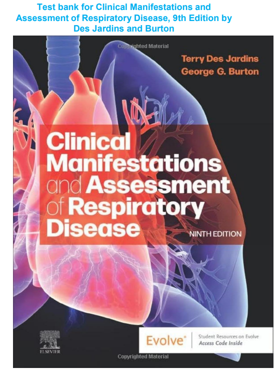
Clinical Manifestations and Assessment of Respiratory Disease by Des Jardins and Burton has long been regarded as one of the most authoritative and clinically grounded texts in respiratory care education. The 9th edition carries that tradition forward with updated content, refined case presentations, and a strong emphasis on connecting assessment findings to pathophysiological mechanisms. It does not simply describe what respiratory diseases look like. It builds the analytical framework clinicians need to understand why those diseases present the way they do and how assessment data guides diagnosis and management. This test bank was written to reinforce and test that same depth of understanding.
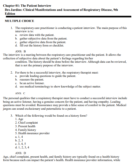
Every question in this resource follows the 9th edition chapter structure. The coverage is comprehensive and clinically focused, moving from the foundational anatomy and physiology of the respiratory system through the mechanics of ventilation, oxygenation and acid-base physiology, and the full spectrum of respiratory diseases encountered in clinical practice. You will find questions covering obstructive lung diseases including chronic obstructive pulmonary disease, asthma, and bronchiectasis, as well as restrictive conditions, pneumonia, tuberculosis, lung cancer, pleural diseases, pulmonary vascular disorders, sleep-disordered breathing, neonatal and pediatric respiratory conditions, and the management of patients requiring mechanical ventilatory support. No major disease category has been omitted.
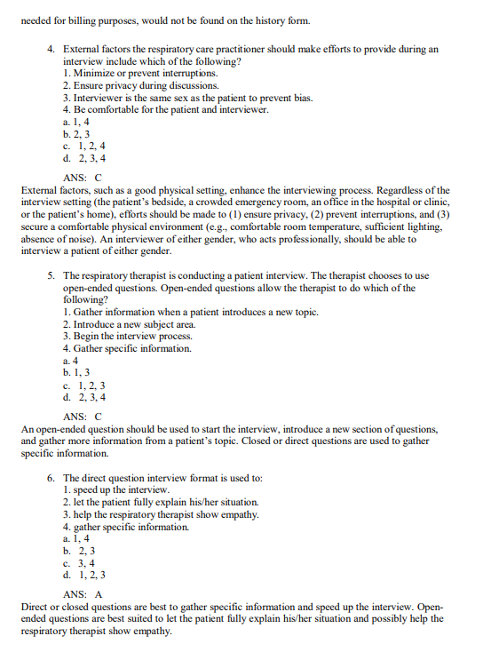
What sets this test bank apart from basic review resources is the quality and depth of its answer explanations. Every question presents a clear clinical scenario, identifies the correct answer, and follows it with a thorough rationale. These rationales do not simply confirm which option is right. They explain the pathophysiological reasoning that makes the correct answer the most accurate choice, address why each of the remaining options is incorrect, and connect the assessment finding or clinical decision back to the underlying disease mechanism. For students in respiratory care and allied health programs where this textbook is a primary resource, that integration of assessment knowledge and pathophysiology is exactly what board examinations and clinical practice demand.
The file is a fully searchable PDF. When you need to focus your review on arterial blood gas interpretation before a clinical competency assessment, or work through the distinguishing features of obstructive versus restrictive patterns before a written examination, use Ctrl+F to navigate directly to that content in seconds. No time lost scrolling through chapters you have already covered. No disruption to your study focus. Precise, targeted access to exactly the material you need right now.
Your download link is generated the moment your purchase is confirmed. No waiting period, no approval process, no account creation required. Open the file on your laptop, tablet, or phone and begin studying immediately. The file is yours permanently with no subscription fee, no renewal requirement, and no expiry date.
What You Get
- Full chapter coverage matched to the 9th edition layout
- Clinically grounded scenario-based multiple-choice questions across all major respiratory disease categories
- Every question answered with the correct response clearly identified
- Detailed rationales explaining the pathophysiology and addressing all answer options
- Searchable PDF format for fast, targeted review by disease, assessment finding, or clinical concept
- One-time purchase with permanent, unlimited file access
Who This Is For
Respiratory therapy students working through their program curriculum will benefit most directly from this resource. It is also highly valuable for students preparing for the Therapist Multiple-Choice and Clinical Simulation examinations administered by the National Board for Respiratory Care, where clinical assessment and disease management are central testing domains. Nursing students who want deeper preparation on respiratory content, allied health students in programs that include respiratory pathophysiology, and practicing clinicians refreshing their knowledge before transitioning into pulmonary or critical care settings will all find this test bank a focused and well-organized study tool.
Sample Questions
Question 1
A respiratory therapist is reviewing the chart of a 58-year-old male patient with a long smoking history. The patient’s pulmonary function test results show a reduced FEV1/FVC ratio, an increased total lung capacity, and an increased residual volume. Breath sounds reveal distant air entry with prolonged expiration and scattered expiratory wheezes. Which condition do these findings most consistently describe?
A) Pulmonary fibrosis
B) Pneumonia
C) Chronic obstructive pulmonary disease
D) Pulmonary embolism
Correct Answer: C
Detailed Explanation:
The constellation of findings in this question is a textbook presentation of chronic obstructive pulmonary disease, specifically emphysema with a chronic bronchitis component. The reduced FEV1/FVC ratio is the defining spirometric feature of obstructive lung disease. In COPD, airflow obstruction results from loss of elastic recoil in emphysema and from airway narrowing and mucus hypersecretion in chronic bronchitis. The increased total lung capacity and residual volume reflect air trapping, which occurs because the loss of elastic recoil allows the lungs to hyperinflate and makes it difficult to fully exhale. The prolonged expiratory phase is a direct consequence of the increased airway resistance and loss of elastic recoil driving airflow out of the lung. The distant breath sounds result from hyperinflation increasing the distance between the lung parenchyma and the chest wall. The expiratory wheezes reflect airflow turbulence through narrowed and partially obstructed airways.
Option A is incorrect because pulmonary fibrosis is a restrictive lung disease. It produces a reduced total lung capacity and residual volume, a normal or elevated FEV1/FVC ratio, and fine inspiratory crackles rather than wheezes. Option B is incorrect because pneumonia produces consolidation findings including bronchial breath sounds, increased tactile fremitus, and dullness to percussion over the affected area, along with systemic signs of infection. Obstructive spirometry patterns are not a feature of uncomplicated pneumonia. Option D is incorrect because pulmonary embolism does not produce obstructive spirometry patterns. It typically presents with sudden dyspnea, pleuritic chest pain, tachycardia, and hemoptysis, with a clinical picture driven by vascular obstruction rather than airway disease.
Question 2
An arterial blood gas drawn on a patient in the emergency department reveals the following results: pH 7.28, PaCO2 58 mmHg, HCO3 26 mEq/L, PaO2 54 mmHg, SaO2 86%. How should the clinician interpret these findings?
A) Metabolic acidosis with hypoxemia
B) Respiratory alkalosis with adequate oxygenation
C) Respiratory acidosis with hypoxemia and no metabolic compensation
D) Compensated metabolic alkalosis with normal oxygenation
Correct Answer: C
Detailed Explanation:
Interpreting arterial blood gases requires a systematic approach. Beginning with the pH, a value of 7.28 falls below the normal range of 7.35 to 7.45, confirming acidosis. The next step is to identify the primary driver of the acidosis by examining the PaCO2 and bicarbonate. The PaCO2 of 58 mmHg is elevated above the normal range of 35 to 45 mmHg, which in the context of acidosis confirms that carbon dioxide retention is the primary cause. This is respiratory acidosis. The bicarbonate level of 26 mEq/L is at the upper limit of normal, which does not represent a meaningful compensatory response. In true compensated respiratory acidosis, the kidneys retain bicarbonate over 48 to 72 hours to buffer the acidosis and bring the pH closer to normal. The near-normal bicarbonate here suggests this is an acute respiratory acidosis without significant renal compensation, which is consistent with an acute respiratory event such as acute exacerbation of COPD, respiratory muscle fatigue, or acute airway obstruction.
The oxygenation component is assessed separately. A PaO2 of 54 mmHg is significantly below the normal range of 80 to 100 mmHg, confirming hypoxemia. An SaO2 of 86% further supports this finding and indicates that the patient is not being adequately oxygenated, which requires immediate intervention.
Option A is incorrect because metabolic acidosis would present with a low bicarbonate level as the primary abnormality, not an elevated PaCO2. Option B is incorrect because the pH is acidotic, not alkalotic, and the PaO2 is critically low, ruling out adequate oxygenation. Option D is incorrect because compensated metabolic alkalosis would show an elevated bicarbonate as the primary finding with a compensatory rise in PaCO2, and the pH would be near normal rather than significantly reduced.
Question 3
A clinician is assessing a patient who presents with sudden onset pleuritic chest pain, dyspnea, and tachycardia following a long-haul flight. The patient has a history of deep vein thrombosis. Chest auscultation reveals clear breath sounds bilaterally. A chest X-ray shows no infiltrates or consolidation. Which diagnosis should be the primary clinical concern, and which initial diagnostic test is most appropriate?
A) Spontaneous pneumothorax; obtain a repeat chest X-ray in lateral decubitus position
B) Pulmonary embolism; obtain a CT pulmonary angiography or ventilation-perfusion scan
C) Acute asthma exacerbation; obtain peak expiratory flow measurement
D) Community-acquired pneumonia; obtain sputum culture and initiate empiric antibiotics
Correct Answer: B
Detailed Explanation:
The clinical picture presented in this question is highly suspicious for pulmonary embolism. The major risk factors are clearly present. Prolonged immobility during a long-haul flight promotes venous stasis in the lower extremities, and a prior history of deep vein thrombosis significantly elevates the risk of recurrent thromboembolic disease. The symptoms of sudden pleuritic chest pain, dyspnea, and tachycardia are classic features of pulmonary embolism. Pleuritic pain results from inflammation of the pleura adjacent to the infarcted lung segment. Dyspnea and tachycardia occur because obstruction of the pulmonary vasculature increases dead space ventilation, impairs gas exchange, and elevates pulmonary vascular resistance, placing acute strain on the right ventricle.
The clear breath sounds and normal chest X-ray are actually consistent with pulmonary embolism. Unlike pneumonia or pneumothorax, pulmonary embolism often produces no abnormality on plain chest radiography in the early stages, or at most subtle findings such as the Westermark sign or Hampton’s hump. CT pulmonary angiography is currently the gold standard imaging modality for diagnosing pulmonary embolism due to its speed, accuracy, and wide availability. Ventilation-perfusion scanning is an appropriate alternative in patients with contraindications to contrast media.
Option A is incorrect because spontaneous pneumothorax would typically present with absent or significantly reduced breath sounds on the affected side and would show a visible pleural line with absence of lung markings on the chest X-ray. Option C is incorrect because asthma typically presents with wheezing, prolonged expiration, and a history of atopy or previous bronchospasm, and the clinical risk factors here point strongly toward a vascular event. Option D is incorrect because community-acquired pneumonia typically presents with fever, productive cough, and consolidation visible on chest X-ray, none of which are present in this scenario.
Question 4
A respiratory therapist is caring for a mechanically ventilated patient in the ICU. The current ventilator settings are assist-control mode with a tidal volume of 700 mL, respiratory rate of 14 breaths per minute, FiO2 of 0.55, and PEEP of 5 cmH2O. The patient weighs 70 kg. The most recent ABG shows pH 7.38, PaCO2 41 mmHg, PaO2 88 mmHg. The peak inspiratory pressure has increased from 28 to 42 cmH2O over the past two hours with no change in plateau pressure. What is the most likely cause of this change?
A) Decreased lung compliance due to worsening pulmonary edema
B) Increased airway resistance due to secretion accumulation or bronchospasm
C) Auto-PEEP caused by an insufficient expiratory time
D) Ventilator circuit disconnection causing air leak
Correct Answer: B
Detailed Explanation:
Understanding the relationship between peak inspiratory pressure and plateau pressure is fundamental to ventilator management and respiratory assessment. Peak inspiratory pressure reflects the total pressure required to deliver the tidal volume and is influenced by both airway resistance and lung compliance. Plateau pressure, measured during an inspiratory hold maneuver, reflects only the elastic recoil of the lung and chest wall and is therefore a measure of static compliance independent of airway resistance.
When peak pressure rises significantly but plateau pressure remains unchanged, the increase in pressure is being generated entirely by increased resistance to airflow through the airways rather than by any change in lung stiffness. The most common causes of a sudden or progressive rise in peak pressure with a stable plateau are secretion accumulation in the endotracheal tube or airways, bronchospasm, biting or kinking of the endotracheal tube, or a mucus plug partially obstructing a major airway. The appropriate response is to suction the airway, assess for bronchospasm and administer bronchodilator therapy if indicated, and inspect the endotracheal tube for obstruction or kinking.
Option A is incorrect because worsening pulmonary edema reduces lung compliance, which would increase plateau pressure alongside peak pressure, not peak pressure alone. Option C is incorrect because auto-PEEP results from insufficient expiratory time allowing incomplete exhalation, which also affects plateau pressure and is associated with increasing baseline airway pressures rather than an isolated rise in peak pressure. Option D is incorrect because a ventilator circuit disconnection or air leak would cause a loss of delivered volume and a decrease in pressure rather than an increase.
Question 5
A clinician is reviewing the chest X-ray of a patient with worsening dyspnea and a dry, nonproductive cough that has been present for several months. The X-ray reveals bilateral interstitial infiltrates with a honeycomb pattern in the lower lobes. Pulmonary function testing shows a reduced total lung capacity, reduced FVC, a normal FEV1/FVC ratio, and reduced diffusing capacity for carbon monoxide. Breath sounds reveal fine, late inspiratory crackles at both lung bases. Which condition do these findings most consistently describe?
A) Chronic obstructive pulmonary disease
B) Idiopathic pulmonary fibrosis
C) Sarcoidosis with upper lobe involvement
D) Hypersensitivity pneumonitis in the acute phase
Correct Answer: B
Detailed Explanation:
The clinical, radiographic, and physiologic findings in this question are classic for idiopathic pulmonary fibrosis, one of the most serious and progressive forms of interstitial lung disease. The bilateral interstitial infiltrates with a honeycomb pattern predominantly in the lower lobes on chest X-ray reflect the usual interstitial pneumonia pattern that is the pathological hallmark of idiopathic pulmonary fibrosis. Honeycombing represents the end-stage fibrotic destruction of normal lung architecture, with formation of cystic airspaces surrounded by dense fibrotic tissue.
The pulmonary function pattern is restrictive, characterized by reduced total lung capacity and FVC with a preserved or elevated FEV1/FVC ratio, reflecting the fact that all lung volumes are reduced proportionally due to the stiff, fibrotic lung tissue rather than the airflow obstruction seen in obstructive diseases. The reduced diffusing capacity for carbon monoxide reflects the destruction of the alveolar-capillary membrane by fibrosis, which impairs gas transfer. The fine, late inspiratory crackles, often described as sounding like Velcro being pulled apart, are a classic auscultatory finding in pulmonary fibrosis and result from the sudden opening of stiff, fibrotic airways during inspiration.
Option A is incorrect because COPD is an obstructive disease producing hyperinflation, a reduced FEV1/FVC ratio, and expiratory wheezes rather than the restrictive pattern and crackles described here. Option C is incorrect because sarcoidosis more commonly involves the upper lobes and hilar lymphadenopathy, and while it can produce a restrictive pattern, the honeycomb pattern in the lower lobes with the specific auscultatory and clinical features described is more consistent with idiopathic pulmonary fibrosis. Option D is incorrect because acute hypersensitivity pneumonitis typically presents with a more abrupt onset following a clear exposure history, and the acute phase produces ground-glass opacities rather than a honeycomb pattern, which represents chronic irreversible fibrosis.
FAQ
Is this an official publisher product?
No. This is an independently written study resource structured around the topics and chapter layout of the 9th edition. It is not affiliated with or endorsed by the authors or their publisher.
Will these questions match my program exams or NBRC board examinations?
This test bank is designed to deepen your understanding of respiratory disease assessment and pathophysiology so you are well prepared for any examination format. It does not preview specific program exams or licensed board examination content.
What makes this test bank different from others?
The detailed rationales go beyond confirming the correct answer. They explain the underlying pathophysiology, connect clinical findings to disease mechanisms, and clarify why each incorrect option fails. That approach builds the kind of deep, transferable understanding that holds up under examination pressure and translates directly into clinical practice.
What file format will I receive?
A fully searchable PDF, navigable by chapter, disease category, assessment finding, or keyword using any standard PDF reader on any device.
How quickly can I access the file after purchase?
Instantly. Your download link is generated immediately after checkout with no waiting period or additional steps required.
Can I open this on my phone or tablet?
Yes. The PDF opens cleanly on any iOS or Android device using a free PDF reader app with no formatting issues.
Do I need the textbook to use this?
Having the textbook alongside is helpful since the chapter order mirrors the 9th edition closely. The questions and rationales are also written in enough detail to be used independently for targeted review and board preparation.
Is there a subscription or renewal fee?
No. This is a one-time purchase. The file is yours to keep and use as many times as you need with no recurring charges or expiry date.

Dorcas M. –
Lots of relevant practice questions with detailed answers and rationales
Lisa Horton –
A solid test bank
Alicia Silva –
Excellent supplement for preparing thoroughly for exams
Warren Brent –
Clear explanations helped me understand challenging topics better.
Terence V. –
Useful test bank with plenty of practice opportunities
Liam Mbogo –
Great resource for reviewing material before upcoming exams
Melania Angel –
Worth every penny for students