The head and neck region is one of the most anatomically complex areas of the human body. Within a relatively compact space, you have bones, muscles, nerves, blood vessels, lymphatic structures, salivary glands, and specialized sensory organs all layered together in intricate relationships. For dental hygienists, dentists, oral health professionals, and students in allied health programs, mastering this anatomy is not optional. It is the foundation on which every clinical skill is built. You cannot administer a nerve block confidently without knowing the course of the inferior alveolar nerve. You cannot interpret a radiograph accurately without understanding the bony landmarks of the skull. You cannot recognize a swollen lymph node without knowing its normal location and drainage territory. This test bank was built to help you develop that depth of understanding.
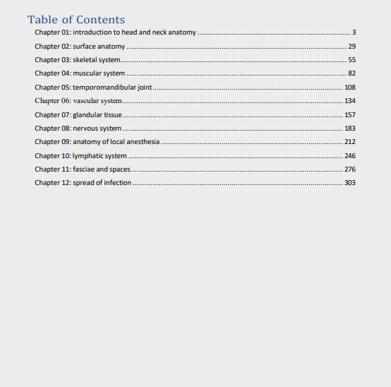
Every question follows the 7th edition chapter structure. The coverage is thorough and logically organized, progressing from the foundational overview of head and neck anatomy through the bones of the skull and face, the muscles of mastication and facial expression, the temporomandibular joint, the major and minor salivary glands, the blood supply and venous drainage of the head and neck, the lymphatic system and associated nodes, and the extensive network of cranial nerves that governs sensation and motor function in this region. No major anatomical area is left uncovered.
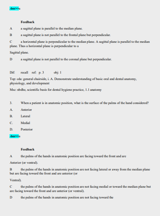
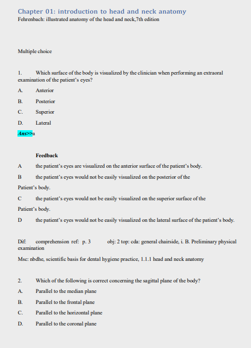
What makes this test bank particularly valuable is the level of detail in its answer explanations. Every question includes the correct answer and a thorough rationale. These explanations do not simply restate the correct choice. They explain the anatomical reasoning behind it, connect the structure to its clinical relevance, and walk through why each incorrect option falls short. For students in dental hygiene, dental assisting, or oral and maxillofacial programs, that connection between anatomy and clinical application is exactly what the board examinations and real practice demand.
The file is a fully searchable PDF. When you need to focus on the branches of the trigeminal nerve before a nerve block practical, or review the contents of the infratemporal fossa before a written examination, use Ctrl+F to navigate directly to that topic. No scrolling through chapters you have already covered. No time lost hunting for the right section. Just precise, efficient studying targeted at exactly what you need to review right now.
Your download link is ready the moment your purchase goes through. No waiting period, no approval process, no account creation required. Open the file on your laptop, tablet, or phone and start working through it immediately. The file is yours permanently, with no subscription and no expiry date.
What You Get
- Full chapter coverage matched to the 7th edition layout
- Scenario and application-based multiple-choice questions across all anatomical regions
- Every question answered with the correct response clearly identified
- Detailed rationales that explain the anatomy and address all answer options
- Searchable PDF format for fast, targeted review by region, structure, or nerve
- One-time purchase with permanent, unlimited file access
Who This Is For
Dental hygiene students working through their anatomy coursework will benefit most directly from this resource. It is also highly valuable for dental assisting students, dental therapy students, and candidates preparing for the National Board Dental Hygiene Examination, where head and neck anatomy is a consistently tested content area. Allied health students in programs that include craniofacial anatomy, speech-language pathology students, and radiography students who need a strong anatomical foundation for interpreting head and neck imaging will also find this test bank a useful and well-organized study tool.
Sample Questions
Question 1
A dental hygiene student is reviewing the bones of the skull and notes that one bone forms the posterior portion of the hard palate. Which bone is being described?
A) Maxilla
B) Palatine bone
C) Vomer
D) Sphenoid bone
Correct Answer: B
Detailed Explanation:
The hard palate is formed by two bones working together. The anterior portion, which makes up roughly two-thirds of the hard palate, is formed by the palatine processes of the maxillae. The posterior portion, making up approximately one-third of the hard palate, is formed by the horizontal plates of the paired palatine bones. This distinction is clinically important in dentistry and dental hygiene because it affects the interpretation of palatal radiographs, the administration of palatal nerve blocks, and the understanding of palatal pathology.
Option A is incorrect because the maxilla contributes to the anterior hard palate, not the posterior portion. Option C is incorrect because the vomer is a thin, flat bone that forms the inferior and posterior part of the nasal septum. It does not contribute to the hard palate. Option D is incorrect because the sphenoid bone contributes to the floor of the cranial cavity and the lateral skull but does not form part of the hard palate. Students frequently confuse the contributions of the maxilla and palatine bones, making this distinction a high-yield topic for board examination preparation.
Question 2
During a patient examination, a dental hygienist palpates a small, firm lymph node located just anterior to the ear at the level of the tragus. Which lymph node is most likely being palpated?
A) Submandibular lymph node
B) Superficial parotid lymph node
C) Jugulodigastric lymph node
D) Submental lymph node
Correct Answer: B
Detailed Explanation:
The superficial parotid lymph nodes, also called preauricular nodes, are located just anterior to the tragus of the ear, overlying or near the parotid gland. These nodes receive lymphatic drainage from the scalp, lateral face, external ear, and eyelids. They drain into the deep parotid nodes and subsequently into the deep cervical chain. Enlarged preauricular nodes can indicate infection or malignancy in their drainage territories, making accurate identification important during extraoral head and neck examinations.
Option A is incorrect because the submandibular lymph nodes are located along the inferior border of the mandible, beneath the chin toward the angle of the jaw, not near the ear. Option C is incorrect because the jugulodigastric node, also called the tonsillar node, is a large node located deep in the neck where the posterior belly of the digastric muscle crosses the internal jugular vein. It is most commonly enlarged during tonsillar infections. Option D is incorrect because the submental nodes are located in the midline beneath the chin and drain the lower lip, chin, and tip of the tongue.
Question 3
A patient presents with inability to open the mouth wide, pain in the preauricular area, and clicking sounds during jaw movement. The clinician suspects involvement of which joint?
A) The atlanto-occipital joint
B) The temporomandibular joint
C) The zygomaticotemporal suture
D) The sphenomandibular articulation
Correct Answer: B
Detailed Explanation:
The temporomandibular joint, or TMJ, is the articulation between the condylar process of the mandible and the mandibular fossa and articular eminence of the temporal bone. It is a synovial joint divided into upper and lower compartments by an articular disc. The signs described in this question, including limited mouth opening, preauricular pain, and clicking or popping sounds during movement, are classic features of temporomandibular disorder, commonly referred to as TMD. Clicking sounds are often associated with disc displacement or abnormal disc movement during condylar translation. Because the TMJ lies just anterior to the external acoustic meatus, pain from this joint is commonly felt in the preauricular region and may radiate to the ear, temple, or jaw.
Option A is incorrect because the atlanto-occipital joint connects the skull to the first cervical vertebra and is responsible for nodding movements of the head. It has no role in jaw opening. Option C is incorrect because the zygomaticotemporal suture is a fibrous joint between the zygomatic bone and temporal bone, not a synovial joint involved in mandibular movement. Option D is incorrect because the sphenomandibular ligament is a ligament associated with the TMJ, not a separate articulation with independent movement.
Question 4
A dental hygienist is preparing to administer an inferior alveolar nerve block. Which foramen must the anesthetic solution be deposited near to achieve anesthesia of the mandibular teeth on that side?
A) Mental foramen
B) Incisive foramen
C) Mandibular foramen
D) Greater palatine foramen
Correct Answer: C
Detailed Explanation:
The inferior alveolar nerve, a branch of the posterior division of the mandibular nerve (V3), enters the mandible through the mandibular foramen, which is located on the medial surface of the ramus of the mandible. To achieve an inferior alveolar nerve block, the clinician deposits local anesthetic just superior and posterior to the mandibular foramen before the nerve enters the bone. Once blocked, this nerve provides anesthesia to the mandibular teeth on the injected side, the lower lip, chin, and buccal gingiva of the anterior teeth via its cutaneous branch, the mental nerve.
Option A is incorrect because the mental foramen is the exit point of the mental nerve on the external surface of the mandibular body, roughly below the premolars. Depositing anesthetic here would only block the mental nerve branches in that area, not the entire inferior alveolar nerve. Option B is incorrect because the incisive foramen is located in the anterior hard palate and is associated with the nasopalatine nerve, which provides palatal sensation to the maxillary anterior teeth. Option D is incorrect because the greater palatine foramen is located in the posterior hard palate and is the target for the greater palatine nerve block, which anesthetizes the posterior hard palate and palatal gingiva of the maxillary posterior teeth.
Question 5
A student is reviewing the muscles of mastication and notes that one muscle originates from the infratemporal crest of the sphenoid bone and the lateral pterygoid plate, and inserts into the medial surface of the mandibular ramus and angle. Which muscle is being described?
A) Masseter
B) Temporalis
C) Medial pterygoid
D) Lateral pterygoid
Correct Answer: C
Detailed Explanation:
The medial pterygoid muscle has a deep head that originates from the medial surface of the lateral pterygoid plate and a superficial head that originates from the pyramidal process of the palatine bone and the maxillary tuberosity. It inserts on the medial surface of the mandibular ramus and angle, forming a muscular sling with the masseter on the opposite side of the ramus. Its primary action is to elevate the mandible, closing the mouth. It also contributes to lateral excursion and protrusion of the mandible. The medial pterygoid is an important landmark in understanding the infratemporal fossa and in clinical procedures such as inferior alveolar nerve blocks, where proximity to this muscle guides needle placement.
Option A is incorrect because the masseter originates from the zygomatic arch and inserts on the lateral surface of the mandibular ramus and angle, not the medial surface. Option B is incorrect because the temporalis originates from the temporal fossa and inserts on the coronoid process of the mandible and the anterior border of the ramus, not the medial ramus surface. Option D is incorrect because the lateral pterygoid has two heads that originate from the greater wing of the sphenoid and the lateral pterygoid plate, and it inserts into the pterygoid fovea of the condyle and the articular disc of the TMJ. Its primary action is to open the mouth and protrude the mandible, not to close it.
(Full answer key with detailed rationales included for all questions in the complete document.)
FAQ
Is this an official publisher product?
No. This is an independently written study resource structured around the topics and chapter layout of the 7th edition. It is not affiliated with or endorsed by the author or their publisher.
Will these questions match my course exams or board examinations?
This test bank is designed to strengthen your understanding of head and neck anatomy and its clinical applications so you are well prepared for any assessment. It does not preview specific course exams or board examination questions.
What makes this test bank different from others?
The detailed rationales go beyond confirming the correct answer. They explain the underlying anatomy, connect it to clinical relevance, and address why each incorrect option is wrong. That approach builds lasting understanding rather than short-term answer recognition.
What file format will I receive?
A fully searchable PDF, navigable by chapter, anatomical region, structure, or nerve using any standard PDF reader on any device.
How quickly can I access the file after purchase?
Instantly. Your download link is generated immediately after checkout with no waiting period or additional steps required.
Can I open this on my phone or tablet?
Yes. The PDF opens cleanly on any iOS or Android device using a free PDF reader app with no formatting issues.
Do I need the textbook to use this?
Having the textbook alongside is helpful since the chapter order mirrors the 7th edition closely. The questions and rationales are also written in enough detail to be used independently for focused review and board preparation.
Is there a subscription or renewal fee?
No. This is a one-time purchase. The file is yours to keep and use as many times as you need with no recurring charges or expiry date.
Melania Angel –
Perfectly organized study guide for exam prep
Norah Helen –
Very good
Winnie M. –
Very useful practice questions
Liam Mbogo –
Great questions and detailed explanations
Mary Kesh –
Boosted my NCLEX confidence significantly
Mary Lincoln –
Very useful study resource
Patty Townsend –
Clear questions with useful answer explanations.
Yunice M –
Excellent resource for exam preparation
Lisa Horton –
Very helpful for testing my knowledge.
Bruno Molino –
Convenient way to review important material.
Caleb Martins –
Good variety of questions with answers and clear explanations.